
Normal Kidney Nuclear Medicine Scan
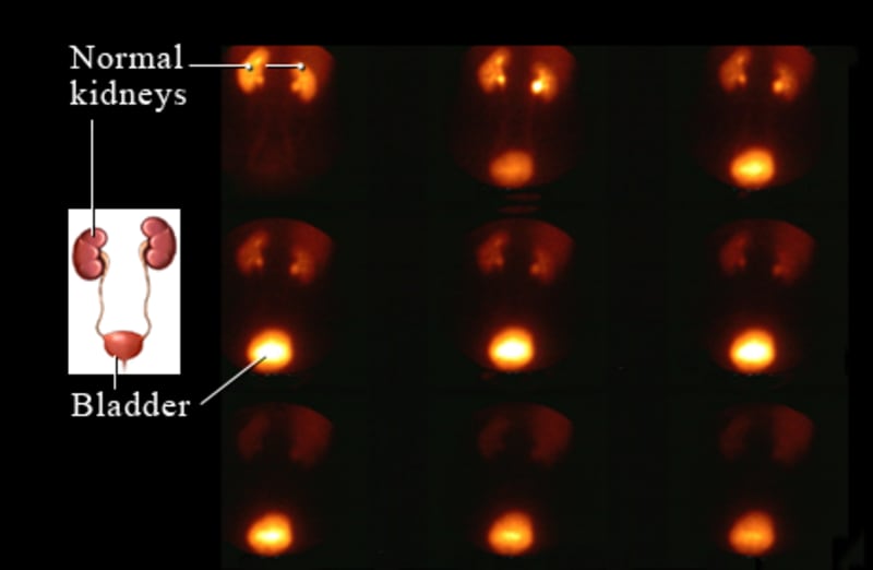
Image courtesy of Intermountain Medical Imaging, Boise, Idaho.
Kidney illustration © Healthwise, Incorporated
This kidney nuclear medicine scan shows the normal flow of radioactive tracer from the kidneys, through the ureters, to the bladder.
Why is a kidney scan done?
A kidney scan is done to:
- Check the blood flow through the kidneys. Abnormal flow may mean narrowed renal arteries that can cause a type of high blood pressure called renovascular hypertension.
- See how a transplanted kidney is working.
- Check the extent of kidney damage caused by an injury or infection.
- Find an obstruction in the kidney or ureter, such as from a kidney stone.
- Find growths in the kidneys (rare).
How is a kidney scan done?
Before the test
You will need to remove any jewelry that might interfere with the scan. You may need to take off all or most of your clothes, depending on which area is being examined. (You may be allowed to keep on your underwear if it does not interfere with the test.) You will be given a cloth or paper covering to use during the test.
You may be asked to drink 2 to 3 glasses of water right before the scan.
During the test
The technologist cleans the site on your arm where the radioactive tracer will be injected. A small amount of the radioactive tracer is then injected. Medicine to increase your urine output (a diuretic) may also be injected. You may lie on your back on a table, stand, or sit upright. A large scanning camera will be positioned closely above your belly.
The camera will scan for radiation right after the radioactive tracer is injected. Scans may be taken every few minutes for about 30 minutes. More pictures may be taken 1 to 2 hours after the tracer was injected. The scans produce pictures as the tracer moves through your kidneys. You may also be given medicine to help the scans check for certain kidney functions.
A chart called a renogram may be made using the information from the kidney scan by plotting the movement of the tracer through the kidneys and recording it on a graph. A series of chart recordings is then made based on the amount of tracer uptake in the kidneys over a period of time. These recordings provide information about different phases of blood flow and kidney function.
You need to remain very still during each scan to avoid blurring the pictures. The camera does not produce any radiation, so you are not exposed to any more radiation while the scan is being done.
How do you prepare for a kidney scan?
If you are breastfeeding, you may want to pump enough breast milk before the test to get through 1 to 2 days of feeding. The radioactive tracer used in this test can get into your breast milk and is not good for the baby.
What do the results of a kidney scan mean?
The results of a kidney scan are usually available in 2 days.
Normal: | The radioactive tracer flows evenly to and through each kidney at the same time. The kidneys are working normally. |
|---|---|
| The tracer flows from the kidneys into the urine, which then drains into the ureters and bladder. This process occurs within a normal time range. | |
| The kidneys take up the radioactive tracer evenly. No "hot" spots or "cold" spots are seen. | |
Abnormal: | The kidneys are not normal in shape, size, or location. |
| The tracer does not flow evenly through the kidneys, meaning narrowing of, blockage of, or damage to the blood vessels or tissue in the kidneys. This may also mean poor kidney function. | |
| The tracer collects in an area ("hot" spot) of a kidney. This might mean a tumor containing a higher-than-normal number of blood vessels. | |
| An area of the kidney does not take up the tracer ("cold" spot). This may mean an abscess, cyst, or scarring. | |
| The tracer does not pass from the kidneys into the urine and then through the ureters to the bladder. This can mean the movement of urine from the kidney is blocked. |
©2011-2026 Healthwise, Incorporated