
What is a magnetic resonance imaging (MRI) of the lumbar spine?
MRI (magnetic resonance imaging) is a test that uses a magnetic field and pulses of radio wave energy to make pictures of the organs and structures inside the body. An MRI can give your doctor information about the spine in your lower back (the lumbar spine). This can include the spine, the space around the spinal cord, and the vertebrae in your lower back.
When you have an MRI, you lie on a table that moves into the MRI machine.
Why is an MRI of the lumbar spine done?
An MRI of the lumbar spine can help find the cause of symptoms like back pain or leg pain, weakness, or numbness. It can help find problems such as a herniated disc, a tumor, or an infection.
Lumbar spine MRI
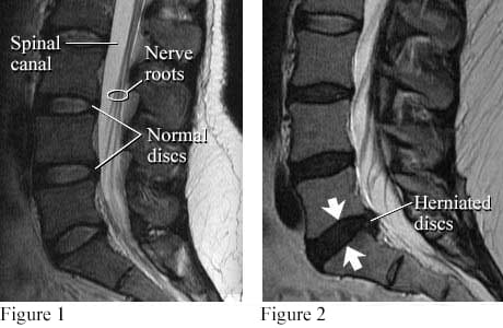
Image courtesy of Intermountain Medical Imaging, Boise, Idaho. All rights reserved.
A side view of the lumbar spine shows normal discs, spinal canal, and nerve roots (see figure 1). Nerve roots normally float in the fluid-filled canal. Figure 2 shows a small herniated disc pushing into the canal toward nerve roots.
©2011-2026 Healthwise, Incorporated